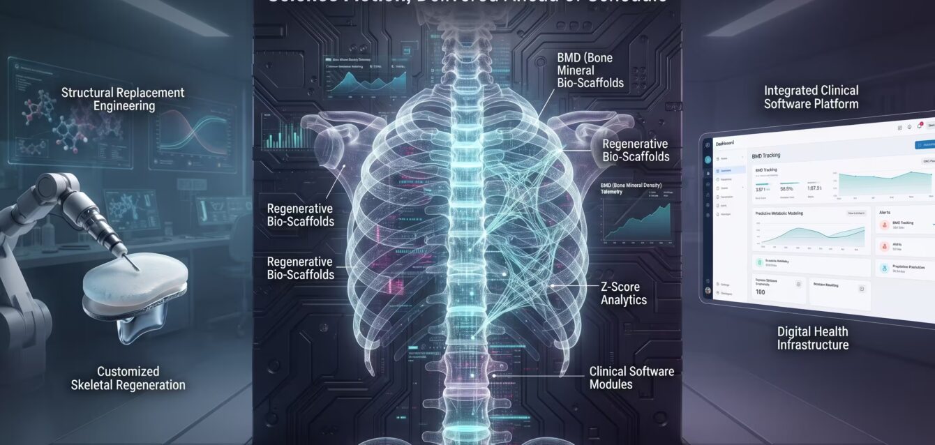
The Bio-Fabricated Skeleton: Science Fiction, Delivered Today
What once lived strictly in the realm of speculative fiction—robotic skeletal reconstruction, engineered bio-scaffolds, and instant structural repair—is rapidly becoming clinical reality. Bridging the gap between legacy medical data and modern bio-engineering, the modern healthcare landscape is transforming how we mend the human body. This deep dive explores the four-stage skeletal regeneration pipeline, examining how advanced data acquisition, custom fabrication, and integrated clinical software are delivering tomorrow’s medical breakthroughs ahead of schedule.
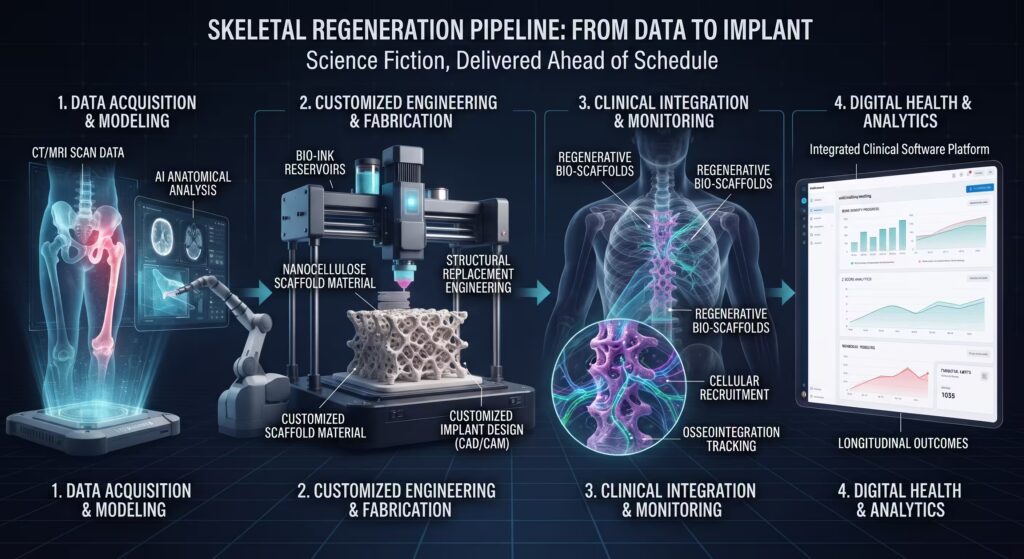
Stage 1: Data Acquisition & AI Modeling
Every breakthrough in modern skeletal regeneration begins with a fundamental transformation in data capture. What was once a labor-intensive, manual process of interpreting flat, two-dimensional radiological slices has evolved into an automated, high-precision pipeline driven by artificial intelligence and volumetric imaging.
1. High-Resolution Volumetric Scanning
The pipeline starts at the diagnostic level. Standard computed tomography (CT) and magnetic resonance imaging (MRI) protocols capture raw volumetric data—often comprising hundreds of axial DICOM slices mapped at sub-millimeter intervals. This raw data preserves fine-grained details regarding bone density, cortical thickness, trabecular architecture, and soft-tissue boundaries.
2. AI-Powered Bone Segmentation
Once the raw scan is ingested, deep learning architectures (such as advanced 3D U-Net and self-configuring segmentation frameworks like nnU-Net) take over. Rather than requiring a clinician to manually trace anatomical borders slice by slice, neural networks classify every voxel in a single forward pass. Within minutes, the system isolates complex structures—accurately separating damaged bone matrices from surrounding vasculature and muscle tissue.
3. Predictive 3D Reconstruction & CAD Readiness
The final output of Stage 1 is an anatomically complete, digital twin of the patient’s skeletal defect. Converted into standard polygonal mesh formats (such as STL files), this volumetric model provides the exact spatial dimensions, structural contours, and density profiles required to feed directly into computer-aided manufacturing systems—setting the stage for custom bio-scaffold fabrication.
“By shifting bone segmentation and volumetric modeling from manual radiology to automated neural networks, we eliminate the latency between diagnosis and precision engineering—turning complex trauma reconstruction into a predictable digital pipeline.”
Stage 2: Customized Bio-Engineering & Fabrication
Once the digital twin and volumetric measurements are finalized, the workflow transitions from computational modeling to physical reality. Stage 2 bridges the gap between raw data and biocompatible hardware through automated computer-aided manufacturing and advanced material science.
1. CAD/CAM and Custom Implant Architecture
Using the precise 3D mesh files generated in Stage 1, computer-aided design (CAD) software allows engineers and clinical specialists to design custom implants tailored down to the micron. Instead of forcing a patient’s anatomy to fit a standard off-the-shelf metal plate or generic bone graft, the implant is engineered to mirror the exact geometry of the missing or damaged bone structure—complete with load-bearing support channels.

2. Advanced Bio-Printing & Nanocellulose Scaffolding
The fabrication process utilizes next-generation 3D bioprinters equipped with specialized bio-ink reservoirs. Rather than relying on rigid, inert titanium or painful autografts that require secondary harvest sites, modern fabrication leverages biocompatible polymers and nanocellulose scaffold materials. These printers deposit material layer-by-layer to build highly porous, interconnected matrix structures designed to act as temporary architectural frameworks for the body.
3. Preparing for Cellular Integration
The ultimate goal of this fabrication phase is not just a structural fix, but a biological one. The intricate porosity of the printed scaffold is engineered specifically to match the mechanical stiffness of natural bone while optimizing surface area—preparing the implant to seamlessly recruit host cells, encourage vascular ingrowth, and facilitate complete tissue regeneration in subsequent stages.
“We are no longer constrained by the limitations of standard shapes and sizes. By pairing precision CAD design with advanced nanocellulose bioprinting, every implant is a bespoke structural solution manufactured precisely for the body it serves.”
Stage 3: Clinical Integration & Bio-Scaffold Functionality
With the bespoke bio-scaffold fabricated and implanted, the focus shifts from engineering and manufacturing to biological assimilation. Stage 3 represents the critical phase where inanimate biomaterial transitions into a living, integrated component of the skeletal system.
1. Cellular Recruitment and Vascular Ingrowth
Once placed within the skeletal defect, the porous nanocellulose or biocompatible ceramic matrix acts as an active biological catalyst. Its interconnected micro-architecture sends biochemical cues that recruit endogenous stem cells, osteoblasts, and endothelial cells from the surrounding host tissue. Concurrently, angiogenesis begins—forming new micro-blood vessels that permeate the scaffold’s internal channels to deliver vital oxygen and nutrients.

2. Osseointegration and Structural Replacement
As host cells populate the scaffold, they begin laying down new extracellular matrix proteins, which subsequently undergo mineralization. Over time, the synthetic or bi-phasic scaffold material gradually degrades at a rate matched by the deposition of native bone. This process ensures true osseointegration, where the boundary between the implant and the host bone dissolves into a continuous, load-bearing structural network.
3. Dynamic Biological Adaptation
Unlike traditional rigid metal plates that can cause stress shielding and weaken surrounding bone, modern regenerative scaffolds are engineered to mirror natural biomechanical elasticity. As mechanical loading resumes through physical movement, the newly formed bone adapts dynamically, thickening and remodeling along lines of stress just like a natural skeleton.
“The true triumph of modern regenerative medicine is not just that the body accepts the implant, but that the implant eventually disappears, replaced entirely by living, breathing bone engineered to the body’s exact specifications.”
Stage 4: Digital Health & Analytics
The final stage of the pipeline shifts focus from the physical implant to the digital nervous system that monitors, models, and optimizes its long-term success. Stage 4 bridges modern digital health infrastructure with the foundational legacy of clinical data systems.
1. Integrated Clinical Software Platforms
Historically, tracking bone mineral accrual and structural integration relied on fragmented, static web calculators—such as the legacy tools once hosted during the early days of pediatric bone density research. Today, those isolated scripts have been replaced by unified, cloud-backed clinical software platforms. These systems ingest longitudinal telemetry from follow-up imaging and patient sensors, maintaining a continuous record of the healing process.

2. Advanced Z-Score Analytics and Predictive Modeling
Modern platforms use sophisticated biostatistical engines to run dynamic Z-score analytics. By comparing real-time post-operative metrics against normalized reference curves, the software calculates exact standard deviations for bone density, structural integrity, and mineral content. Furthermore, machine learning models analyze these trajectories to predict long-term integration outcomes—flagging potential anomalies or delayed ossification months before they manifest physically.
3. Closing the Loop
By combining high-precision data acquisition, automated bioprinting, biological assimilation, and predictive analytics, the modern skeletal regeneration pipeline is fully closed. It transforms medical intervention from an isolated event into a continuous, data-driven science—proving once again that yesterday’s speculative fiction is today’s functional infrastructure.
“The future of medicine isn’t just about replacing what is broken; it’s about building a continuous digital feedback loop that ensures the body heals smarter, faster, and with absolute precision.”
Summary
As we examine the complete pipeline—from AI-driven volumetric modeling and precision nanocellulose bioprinting to biological osseointegration and predictive digital analytics—the trajectory of modern medicine becomes clear. What was once confined to the pages of speculative fiction is now active clinical infrastructure. By bridging historical data frameworks with next-generation digital health systems, we are no longer just treating skeletal trauma; we are engineering solutions that are delivered well ahead of schedule.
Signing off on today’s leap in modern medical advancements brought on by AI and the advanced technologies it supports.